![]() Click to view article in PDF format.
Click to view article in PDF format.
A New Quantitative Method for Analysis of Drill Cuttings and Core for Geologic, Diagenetic and Reservoir Evaluation*
![]() Jon
Jon![]() Sliwinski1, Michael Le Strat2, and Murray Dublonko2
Sliwinski1, Michael Le Strat2, and Murray Dublonko2
Search and Discovery Article #40482 (2010)
Posted February 12, 2010
* Adapted from expanded abstract prepared for AAPG Annual Convention and Exhibition, Denver, Colorado, USA, June 7-10, 2009.
1SGS Minerals Services, Lakefield, ON, Canada ([email protected])
2Breaker Energy Ltd., Calgary, AB, Canada
Drill cuttings have traditionally been utilized as a first look, qualitative tool but are often not re-examined once wireline logs are run. The advent of advanced technology from the mining industry has broad applications for quantitative rock analysis of oil and gas reservoirs. The automated, high resolution data provided from QEMSCAN® analysis of drill cuttings provides much more information than conventional optical analysis.
SGS and Breaker Energy have completed a pilot study to evaluate the applications of this new technique on a recently drilled well. The horizontal well in NE British Columbia targeted the Doig Formation, a tight, shaly gas sand with localized phosphatic zones. The QEMSCAN® data provided mineralogical and textural information on the distribution of coarse grains and shale, visual confirmation of cementation mineralogy and fabric as well as quantitative porosity numbers. The analysis aided in developing the depositional model, digenetic history, reservoir characteristics and completion/production strategies. This paper will present the methodology, data and conclusions of this study.
uAbstract |
Drill cuttings are typically the first piece of data available from the subsurface during drilling. These samples of the subsurface formations are collected at surface and used by wellsite geologists to prepare a striplog and then typically disappear from the data stream. These samples can provide valuable information and in the absence of core provide the only actual contact with the rock. Recently there has been renewed interest in the data available from drill cuttings and the application of advanced techniques (Scanning Electron Microscopy, X-ray Dispersive Energy Spectroscopy, Back Scattered Electron Imaging and Digital Imaging and Processing) as well as advanced sample preparation (grain mount thin sections).
QEMSCAN® is an automated mineralogy analysis system that has a long history in the mining sector. It is a combination of a Scanning Electron Microscope (SEM) and Electron Dispersive Spectroscopy (EDS) with automated analysis software that uses BSE (Backscattered Electron Intensity) and EDX (Energy Dispersive X-ray) spectra to identify the mineral or phase present at the analysis point. QEMSCAN® can measure and identify up to 600 points per second. This speed allows for detailed mineral mapping of samples, with the production of digital images, and superb statistical data on the modal mineralogy; typically millions of data points are measured during each analysis. QEMSCAN® is able to analyze cuttings, as well as conventional and rotary cores. Prepared samples are polished sections from cuttings, polished thin sections from cuttings or core, and solid core.
Drill cuttings were provided from the wellsite and first examined and photographed using a traditional stereoscopic microscope (Figure 1). The wellsite geologist also provided his observations in a traditional striplog for comparison. Polished sections were prepared using micro-riffled samples from each five meter depth interval. The riffling process ensures that a random selection of particles from the interval is present in the analysis. These particles are then prepared in a resin polished section for analysis by QEMSCAN®.
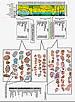
When analyzing samples using QEMSCAN®, there are several choices for operation mode (Figure 2), resolution and magnification. There are several scanning modes depending on what information is sought, for the purposes of this study we ran a line scan (Bulk Modal Analysis) for overall mineralogy and a particle scan (Particle Map Analysis) based on the use of cuttings as opposed to core or thin section. In the mining industry, samples are usually crushed and screened into size fractions and the resolution and magnification is dependent on the size of the fraction being analyzed. In this case, we did not screen the sample as the particle size was fairly consistent at 1-0.5 mm, which also determined the magnification of 20x in order to capture the particles in the view window. The matter of resolution was not as easily determined and several different resolutions were compared. In the end, it was determined that a resolution of 5μm was optimal for showing details of the grains and giving a clear view of the porosity and cementing features. In order to save time in the analysis all particles less than 250 μm were not analyzed. As a control the modal mineralogy from the BMA, run on the entire sample, was compared to the mineralogy from the PMA to ensure the analysis was statistically accurate.
Presented in Figure 3 is the data from one polished section prepared from a depth interval in the Doig Formation. The first image shows the total particles scanned. In this case, there were 197 particles larger than the lower limit of 250 μm.
The second image is one of the sandstone grains enlarged showing the detail. In the false colour map, pink represents quartz, orange potassium feldspar, red pyrite and turquoise dolomite. The grain is very clearly cemented with a dolomitic cement as well the pore space, the white voids, are lined with dolomite suggesting the formation may respond well to an acid treatment prior to the planned frac. Porosity of this particle can be determined by the analysis software and combined with that of the other grains giving a quantitative value for the porosity.
The third image is the individual quartz and potassium feldspar grains from the sandstone which were then analyzed by the software for roundness (shape factor), and average grain size. This textural information can then be incorporated in a depositional model.
The fourth image is the pore throats of the sandstone grain. The analysis software determined the porosity to be 5.06% and have an average size of 10.33 μm. A new porosity value can then be calculated using a minimum “effective” pore size cutoff, giving a value for effective porosity. Alternately part of the dolomitic pore lining could be added to the porosity, simulating an acid stimulation, giving an “improved” porosity value.
The QEMSCAN® provided useful data in the evaluation of this reservoir. The speed and capacity to evaluate large volumes of samples from this horizontal well in a small amount of time, allowed a detailed analysis of small changes within the reservoir. Based on data from the QEMSCAN® analysis it became apparent there were three separate sands intersected by the well bore. Each of these sands had its own distinct mineralogy and textural characteristics as shown in Figure 4.
QEMSCAN® demonstrated it is a valuable new tool to the oil and gas industry. It has broad applications from simple analysis of cuttings for modal mineralogy to detailed mapping of core. It has applications in the analysis of individual wells as well as showing potential for large field studies and field-to-field comparisons.
The authors would like to acknowledge SGS and Breaker Energy for providing the resources to complete this study.
Copyright © AAPG. Serial rights given by author. For all other rights contact author directly. |