Clikc on image for enlargement.
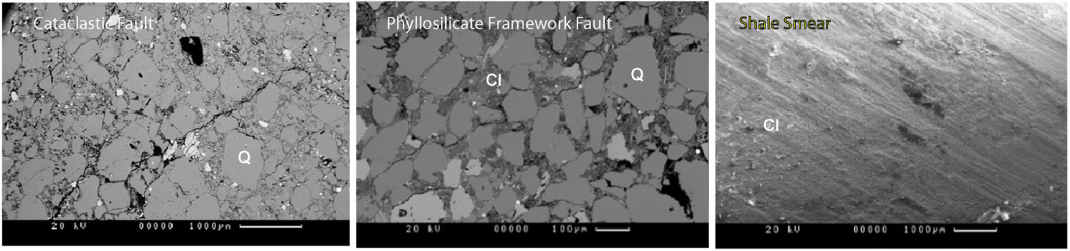
Figure 4. Scanning electron microscope (SEM) images of, from left to right: a low clay-content cataclastic fault rock, a higher clay content mixing of clays with quartz in the phyllosilicate framework fault rock, and a shale smear.