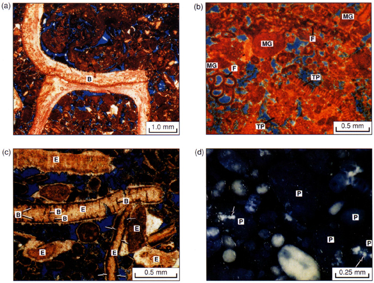
Figure 11 a,b,c,d. Microporous grains in the Jurassic and in the Recent. Porosity in thin section is filled by blue-dyed epoxy, calcite is stained pink to red, other minerals are unstained. (a) Thin section photomicrograph of ooid-skeletal mud-lean packstone. Large brachiopod fragment (B) was originally composed of low magnesium calcite and is non-microporous. In contrast, surrounding grains are micritized and microporous (plane-polarized light, Saudi Arabia, Khursaniyah field, Arab-D). (b) Thin section photomicrograph showing micritized and microporous grains originally composed of metastable high magnesium calcite. Original high magnesium calcite components include several types of foraminifera (F), micritized grains (MG) and the red algae Thaumatoporella (TP) (plane-polarized light, Saudi Arabia, Ghawar field, Arab-D). (c) Thin section photomicrograph showing preserved microstructure of several echinoderm grains (E). In spite of their original high magnesium calcite mineralogy, echinoderms are rarely microporous. Algal or fungal borings (B) and micrite envelopes (arrows) are locally common on these grains, although these grains never appear completely micritized (plane-polarized light, Saudi Arabia, Ghawar field, Arab-D). (d) Photomicrograph of a Recent skeletalpeloidal sand. Peloids (P) are completely micritized and microporous, as indicated by their bluish color. Blue appearance results from the infiltration of blue-dyed epoxy into micropores in the grains. In contrast, solid grains are pearly white in color. Note that some grains are incompletely micritized (arrows), and grade from bluish micritized and microporous CaCO3 into white, solid CaCO3 within the same grain (reflected light, Holocene, North Caicos, British West Indies).